Vision - Structures of the Eye
- The cornea is the transparent front part of the eye that covers the iris, pupil, and anterior chamber.
- The sclera, also known as the white of the eye, is the opaque, fibrous, protective, outer layer of the eye containing collagen and elastic fiber.
- The choroid is the vascular layer of the eye, containing connective tissue, and lying between the retina and the sclera.
- The anterior chamber is the fluid-filled space inside the eye between the iris and the cornea's innermost surface.
- The posterior chamber is a narrow space behind the peripheral part of the iris, and in front of the suspensory ligament of the lens and the ciliary processes.
- The iris is a thin, circular structure in the eye, responsible for controlling the diameter and size of the pupil and thus the amount of light reaching the retina.
- The iris dilator muscle is a smooth muscle of the eye, running radially in the iris and therefore fit as a dilator.
- The iris sphincter muscle encircles the pupil of the iris, appropriate to its function as a constrictor of the pupil.
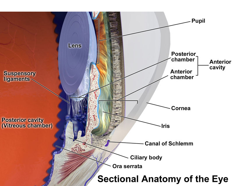
- The ciliary body is a part of the eye that includes the ciliary muscle, which controls the shape of the lens, and the ciliary epithelium, which produces the aqueous humor.
- The aqueous humour is a transparent, gelatinous fluid similar to plasma located in the anterior and posterior chambers of the eye.
- The canal of Schlemm is a circular lymphatic-like vessel in the eye that collects aqueous humor from the anterior chamber and delivers it into the episcleral blood vessels via aqueous veins.
- The crystalline lens is a transparent, biconvex structure in the eye that, along with the cornea, helps to refract light to be focused on the retina.
- The ciliary muscle in the eye's middle layer (vascular layer) that controls accommodation for viewing objects at varying distances and regulates the flow of aqueous humour into Schlemm's canal.
- The vitreous humour is the clear gel that fills the space between the lens and the retina of the eyeball of humans and other vertebrates.

Anatomy of the eye.
