Interdisciplinary Note (6 of 36)
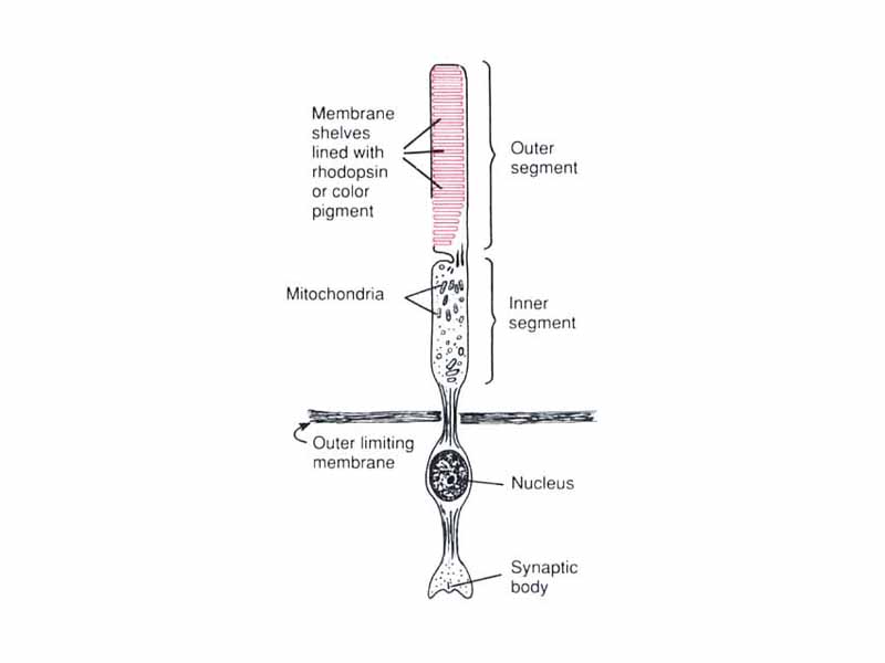
It is helpful to understand that the retina is really part of the brain. The human retina contains two kinds of photoreceptors, rods and cones. Rods and cones both possess opsins, visual pigments formed by the combination of the lipoprotein opsin and the organic pigment retinal, a derivative of vitamin A. Rhodopsin is the type of opsin found in rods. Cones contain a variety of other opsins, called photopsins. Retinal is a very unusual pigment. The absorption of light causes the rotation of a π bond, converting 11-cis retinal into 11-trans retinal. 11-trans retinal doesn't fit well in opsin, triggering changes in the protein conformation. The opsin is now said to be activated. Activated opsin triggers the activation of the signal coupling protein transducin, beginning an amplification cascade similar to a second messenger system (1 activated rhodopsin triggers 500 activated transducin molecules). Activated transducin binds and activates phosphodiesterase (PDE) which then hydrolyzes cGMP to remove it from Na+ channels. One photon leads to the blockage of about 1 million sodium ions, leading to hyperpolarization of the visual receptor cell, which inhibits the spontaneous release of neurotransmitter. Eventually the retinal and opsin dissociate and the retinal is enzymatically reconverted to cis retinal.
