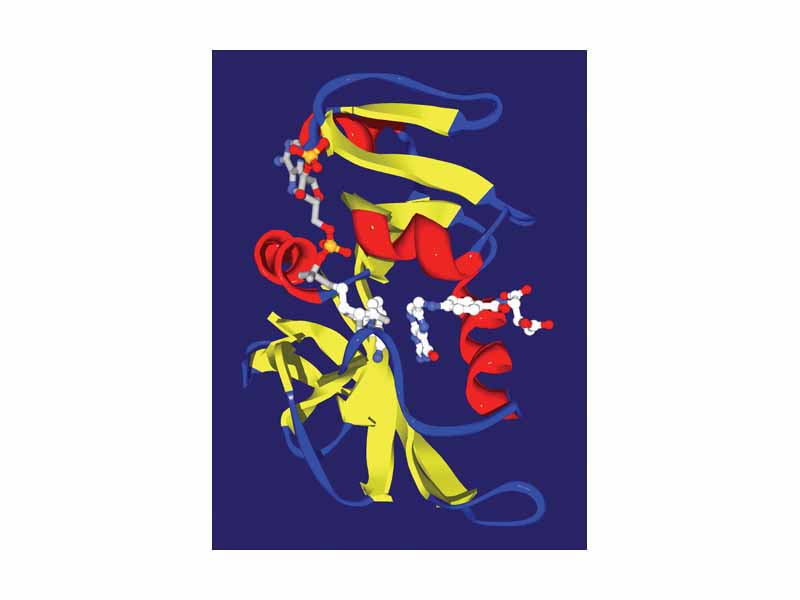
Dihydrofolate reductase from E. coli with its two substrates, dihydrofolate (right) and NADPH (left), bound in the active site. The protein is shown as a ribbon diagram, with alpha helices in red, beta sheets in yellow and loops in blue.
Click this LINK to visit the original image and attribution information. Right click on the image to save the 800px teaching JPEG.
